Mitral Stenosis
Medically reviewed by Drugs.com. Last updated on Sep 1, 2024.
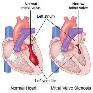
What is mitral stenosis?
Mitral stenosis is a condition that makes your mitral valve narrow and stiff. The mitral valve is between the left atrium and the left ventricle of your heart. The valve opens and closes to direct blood flow through your heart. With mitral stenosis, your valve may not open or close properly. This causes strain on your heart muscle and decreases blood flow to your body.
 |
What causes mitral stenosis?
- Rheumatic fever can develop after you have a strep throat infection. Rheumatic fever can cause inflammation and damage to your mitral valve. The walls of your mitral valve may narrow, and may even join together.
- Calcium buildup can happen as you age. Calcium can build up on the mitral valve walls and make them narrow and stiff.
- Congenital heart defect means you are born with a damaged mitral valve that leads to narrowing and blockage.
What are the signs and symptoms of mitral stenosis?
You may not have any symptoms, if your mitral stenosis is mild. You may have any of the following if it is more severe:
- Shortness of breath during activity or when you lie down
- Severe tiredness
- Swollen feet or ankles
- Fast, jumpy, or fluttery heartbeat
- Coughing up bloody tinged mucus
- Dizziness or fainting
- Headache and stroke symptoms
How is mitral stenosis diagnosed?
Your healthcare provider will ask about your signs and symptoms and listen to your heart. He or she will ask if you have ever had strep throat or rheumatic fever. Tell him or her if you have a family history of heart disease. You may also need any of the following:
- Blood tests may show an infection or other cause of mitral stenosis.
- An echocardiogram is a type of ultrasound. It is used to show problems with your mitral valve and how blood flows through your heart. It may also show how well your heart is pumping. You may need a transthoracic or transesophageal echocardiogram. Ask your healthcare provider about these types of echocardiogram.
- A chest x-ray shows the size of your heart. It may also show if fluid is around your heart and lungs.
- An electrocardiogram (also called an EKG or ECG) is a test that measures the electrical activity of your heart. It is used to show your heart rate and rhythm, and to show how well your heart is working. It may also help your healthcare provider diagnose heart problems.
- A stress test may show the changes that take place in your heart while it is under stress. Stress may be placed on your heart with exercise or medicine. Ask for more information about this test.
- Cardiac catheterization is a procedure to check how well your heart is pumping blood. It is also used to measure pressure in different parts of your heart. A catheter (long thin tube) is inserted into your arm, neck, or groin and moved into your heart. An x-ray may be used to guide the tube to the right place. Contrast liquid may be used to help your heart show up better in the pictures. Tell the healthcare provider if you have ever had an allergic reaction to contrast liquid.
How is mitral stenosis treated?
Treatment for mitral stenosis depends on how severe your symptoms are. If you do not have symptoms, your healthcare provider will do tests regularly. You may also need any of the following if your symptoms become worse:
- Medicines may be used to remove extra fluid, treat arrhythmias (irregular heartbeats), control heart rhythm, or prevent blood clots.
- Balloon valvuloplasty helps widen your mitral valve and allow blood to flow through easier. It is also called a closed valvotomy. A catheter with a balloon on the tip is inserted through a small incision in your arm or groin. The catheter is guided through a blood vessel and into your left atrium near your mitral valve. When the balloon is inflated, it stretches the valve opening.
- Commissurotomy is open heart surgery to fix your mitral valve. It is done if valvuloplasty does not correct your mitral stenosis. During a commissurotomy, your surgeon will remove calcium buildup and scar tissue from your valve.
- Valve replacement is a surgery to remove part or all of your mitral valve. A new valve is then secured in place. The new valve may be from a donor (another person or animal), or may be an artificial valve. There are 2 different approaches for valve replacement. The first is an open heart procedure. The second is a procedure that replaces the valve through a catheter guided into a vessel in your groin. Your healthcare provider will talk to you about which approach is right for you.
How can I manage mitral stenosis?
- Eat heart-healthy foods. Heart-healthy foods include salmon, tuna, walnuts, whole-grain breads, low-fat dairy products, beans, and oils such as olive or canola oil. A dietitian or your provider can give you more information on meal plans such as the DASH (Dietary Approaches to Stop Hypertension) eating plan. The DASH plan is low in sodium, processed sugar, unhealthy fats, and total fat. It is high in potassium, calcium, and fiber. These can be found in vegetables, fruit, and whole-grain foods.

- Limit sodium (salt) as directed. Too much sodium can affect your fluid balance. Check labels to find low-sodium or no-salt-added foods. You can also make small changes to get less salt. For example, if you add salt while you cook, do not add more salt at the table. Ask your healthcare provider or dietitian for more ways to cut down on salt.

- Exercise as directed. Exercise will improve your heart health. Ask your healthcare provider about the best exercise plan for you. Start slowly and increase activity as you get stronger. Stop if you feel short of breath.
- Limit caffeine. Caffeine can make irregular heartbeats worse. Ask your healthcare provider about eating or drinking anything that contains caffeine. Ask him or her how much caffeine is safe for you.
- Do not smoke. Nicotine and other chemicals in cigarettes and cigars can cause lung and heart damage. Ask your healthcare provider for information if you currently smoke and need help to quit. E-cigarettes or smokeless tobacco still contain nicotine. Talk to your healthcare provider before you use these products.
- Limit or do not drink alcohol. Ask your healthcare provider if it is okay for you to drink alcohol. Alcohol can increase your risk for high blood pressure and coronary artery disease. Your provider can tell you how many drinks are okay to have within 24 hours or within 1 week. A drink of alcohol is 12 ounces of beer, 5 ounces of wine, or 1½ ounces of liquor.
- Talk to your healthcare provider about pregnancy. If you are a woman and want to get pregnant, talk to your healthcare provider. You and your baby may need to be monitored by specialists during your pregnancy.
- Ask about vaccines you may need. Certain diseases are dangerous for a person who has mitral stenosis. Vaccines help lower your risk for infections that can lead to disease. Get a flu vaccine as soon as recommended each year, usually in September or October. Your healthcare provider can tell you if you also need other vaccines, and when to get them.
What can I do to prevent mitral stenosis?
- Manage other health conditions. High blood pressure and high cholesterol levels can worsen mitral stenosis. Ask your healthcare provider for more information on managing these or other health conditions.
- Get treatment for strep throat. Strep throat that is not treated can cause rheumatic fever.
Call your local emergency number (911 in the US) or have someone call if:
- You have any of the following signs of a stroke:
- Numbness or drooping on one side of your face
- Weakness in an arm or leg
- Confusion or difficulty speaking
- Dizziness, a severe headache, or vision loss
- You have any of the following signs of a heart attack:
- Squeezing, pressure, or pain in your chest
- You may also have any of the following:
- Discomfort or pain in your back, neck, jaw, stomach, or arm
- Shortness of breath
- Nausea or vomiting
- Lightheadedness or a sudden cold sweat
- You feel lightheaded, short of breath, and have chest pain.
- You cough up blood.
When should I seek immediate care?
- Your arm or leg feels warm, tender, and painful. It may look swollen and red.
- Your symptoms get worse.
When should I call my doctor?
- The veins in your neck look swollen or are bulging.
- You have a fever.
- You have questions or concerns about your condition or care.
Care Agreement
You have the right to help plan your care. Learn about your health condition and how it may be treated. Discuss treatment options with your healthcare providers to decide what care you want to receive. You always have the right to refuse treatment. The above information is an educational aid only. It is not intended as medical advice for individual conditions or treatments. Talk to your doctor, nurse or pharmacist before following any medical regimen to see if it is safe and effective for you.© Copyright Merative 2024 Information is for End User's use only and may not be sold, redistributed or otherwise used for commercial purposes.
Learn more about Mitral Stenosis
Treatment options
Care guides
Further information
Always consult your healthcare provider to ensure the information displayed on this page applies to your personal circumstances.