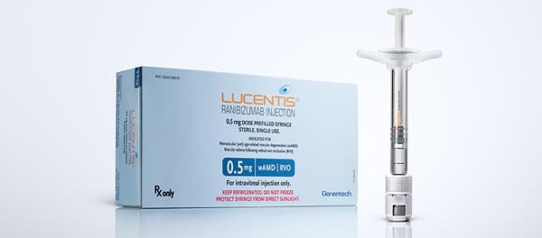
Ranibizumab (Monograph)
Brand name: Lucentis
Drug class: EENT Drugs, Miscellaneous
VA class: OP900
Chemical name: Disulfide with human-mouse monoclonal rhuFAB V2 light chain anti-(human vascular endothelial growth factor) Fab fragment (human-mouse monoclonal rhuFAB V2 γ1-chain) immunoglobulin G1
Molecular formula: C2158H3282N562O681 S12
CAS number: 347396-82-1
Introduction
Recombinant humanized immunoglobulin G1 kappa (IgG1 kappa) monoclonal antibody fragment; a vascular endothelial growth factor A (VEGF-A) antagonist.1 3 4 5 6
Uses for Ranibizumab
Neovascular Age-related Macular Degeneration
Treatment of neovascular (wet) age-related macular degeneration.1 4 5 6 14 19
Macular Edema Following Retinal Vein Occlusion
Treatment of macular edema following retinal vein occlusion.1 11 12 16 18
Diabetic Macular Edema
Treatment of diabetic macular edema.1 12 13 20
Diabetic Retinopathy in Patients with Diabetic Macular Edema
Treatment of diabetic retinopathy (proliferative or nonproliferative) in patients with diabetic macular edema.1 4 13 15
Ranibizumab Dosage and Administration
Administration
Ophthalmic Administration
Administer by intravitreal injection only into the affected eye(s).1
Commercially available as single-use vials containing 0.3 or 0.5 mg of the drug for intravitreal injection.1 Prior to intravitreal administration, withdraw entire contents of the appropriate strength ranibizumab vial a sterile 5-µm, 19-gauge filter needle (provided by manufacturer) into a 1-mL tuberculin syringe using aseptic technique.1 4 Next, replace filter needle with a sterile 30-gauge, ½-inch needle (provided by manufacturer) for intravitreal injection.1 To obtain appropriate dose (0.3 or 0.5 mg), expel contents in tuberculin syringe until plunger tip is aligned with the line that marks 0.05 mL on the syringe.1
Inject under controlled aseptic conditions (including use of sterile gloves, sterile drape, a sterile eyelid speculum [or equivalent]) following adequate anesthesia and administration of a broad-spectrum anti-infective agent.1
Monitor patients for elevation of IOP prior to and 30 minutes following intravitreal injection using tonometry.1 Monitoring may include evaluation of optic nerve head perfusion immediately after injection.1 Monitor patients for any manifestations of endophthalmitis.1 (See Advice to Patients.)
Each vial should be used only for treatment of a single eye.1 If contralateral eye requires treatment, use a new vial; change sterile field, syringe, gloves, drape, eyelid speculum, and filter and injection needles before administering to the other eye.1
Dosage
Adults
Neovascular Age-related Macular Degeneration
Ophthalmic
Intravitreal injection: 0.5 mg (0.05 mL of a solution containing 10 mg/mL) into the affected eye(s) once every month (approximately every 28 days).1
After the first several months of therapy, may reduce dosing frequency to decrease treatment burden; however, less frequent dosing regimens are not as effective as continuous monthly dosing and patients should be evaluated regularly.1 14 19
After the first 3 monthly injections, may reduce to as-needed dosing with regular clinical assessment; while a regimen averaging 4–5 injections over the following 9 months is expected to maintain visual acuity, continuous monthly dosing can result in additional gains (by 1–2 letters).1
After the first 4 monthly injections, may reduce dosing frequency to one injection every 3 months; compared with continuous monthly dosing, such a regimen has been shown to result in an approximate 5-letter (1-line) loss of visual acuity over 9 months.1
Macular Edema Following Retinal Vein Occlusion
Ophthalmic
Intravitreal injection: 0.5 mg (0.05 mL of a solution containing 10 mg/mL) into the affected eye(s) once every month (approximately every 28 days).1
Diabetic Macular Edema
Ophthalmic
Intravitreal injection: 0.3 mg (0.05 mL of a solution containing 6 mg/mL) into the affected eye(s) once every month (approximately every 28 days).1
Diabetic Retinopathy in Patients with Diabetic Macular Edema
Ophthalmic
Intravitreal injection: 0.3 mg (0.05 mL of a solution containing 6 mg/mL) into the affected eye(s) once every month (approximately every 28 days).1
Special Populations
Hepatic Impairment
No specific dosage recommendations at this time.1
Renal Impairment
No dosage adjustment required.1 (See Renal Impairment under Cautions.)
Geriatric Patients
No specific dosage recommendations at this time.1
Related/similar drugs
Syfovre, triamcinolone ophthalmic, dexamethasone ophthalmic, Eylea, Izervay, fluocinolone ophthalmic, Ozurdex
Cautions for Ranibizumab
Contraindications
-
Ocular or periocular infections.1
-
Known hypersensitivity (e.g., severe intraocular inflammation) to ranibizumab or any ingredient in the formulation.1
Warnings/Precautions
Endophthalmitis and Other Serious Ocular Effects
Intravitreal injections, including those with ranibizumab, associated with endophthalmitis and retinal detachments.1 4 5 6 Always use proper aseptic injection technique.1 4 (See Ophthalmic Administration under Dosage and Administration.) Monitor patients closely for signs of endophthalmitis (e.g., redness, sensitivity to light, pain, changes in vision) during the week following injection to permit early treatment.1 4 (See Advice to Patients.)
Traumatic cataract and tearing of retinal pigment epithelium also reported.1 4 5
Increased IOP
Increased IOP observed both before and after (60 minutes after) intravitreal injection.1 4 6 Monitor IOP and perfusion of optic nerve head and manage appropriately.1 4
Thromboembolic Events
Potential risk of arterial thromboembolic events following intravitreal injection of VEGF antagonists.1 Arterial thromboembolic events (i.e., nonfatal stroke, nonfatal MI, vascular death [including deaths from unknown causes]) reported at low rates (0.8–10.8%).1 4 20 Patients with diabetic macular edema and diabetic retinopathy had higher rates, but were typical of this patient population.1 20
Potentially higher rate of stroke associated with the 0.5- versus 0.3-mg dose in patients with neovascular age-related macular degeneration; additional postmarketing surveillance and clinical studies are being conducted to further evaluate this finding.4 7 8 10 17
Fatal Events
In clinical studies in patients with diabetic macular edema and diabetic retinopathy, fatal events occurred more frequently with ranibizumab than sham treatment.1 4 Although the fatality rate was low and included causes typical of patients with advanced diabetic complications, a potential relationship to the drug cannot be excluded.1
Immunogenicity
Development of anti-ranibizumab antibodies reported.1 Clinical relevance unclear, but iritis or vitritis noted in some patients with neovascular age-related macular degeneration who had the highest levels of immunoreactivity.1
Specific Populations
Pregnancy
Category C.1
Studies not conducted in pregnant women; it is not known whether ranibizumab can cause fetal harm when administered during pregnancy.1 Use only if potential benefits justify potential risk to fetus.1 Adverse effects on embryofetal development or reproductive capacity possible due to the drug's mechanism as a VEGF antagonist.1
Lactation
Not known whether ranibizumab is distributed into milk.1 Caution if used in nursing women.1
Pediatric Use
Safety and efficacy not established.1
Adult Use
Safety and efficacy not established in adults <50 years of age.4
Geriatric Use
No substantial differences in efficacy or systemic exposure (after correcting for Clcr) relative to younger adults.1
Hepatic Impairment
Pharmacokinetics not studied; dosage adjustment not expected to be necessary.1
Renal Impairment
Increased ranibizumab exposure observed in patients with renal impairment; however, changes not considered to be clinically important.1 (See Renal Impairment under Dosage and Administration.)
Common Adverse Effects
Conjunctival hemorrhage,1 4 eye pain,1 4 vitreous floaters,1 4 increased IOP,1 4 intraocular inflammation.1 4
Drug Interactions
No formal drug interaction studies to date.1
Photodynamic Therapy with Verteporfin
Serious intraocular inflammation reported; most cases occurred when ranibizumab was administered approximately 7 days after verteporfin photodynamic therapy in patients with neovascular age-related macular degeneration.1
Ranibizumab Pharmacokinetics
Absorption
Bioavailability
Following monthly intravitreal injection, peak serum concentrations attained were substantially below that necessary to inhibit the biologic activity of VEGF-A by 50%.1 Serum concentrations predicted to be approximately 90,000 times lower than vitreal concentrations.1
Peak serum concentrations predicted to be reached approximately 1 day after monthly intravitreal administration of 0.5 mg per eye.1
Elimination
Half-Life
Estimated average vitreous half-life: Approximately 9 days.1
Stability
Storage
Ophthalmic
2–8°C.1 Do not freeze; protect from light.1 Store in original carton until use.1
Actions
-
Binds to active forms of human VEGF-A, including cleaved form (VEGF110), and inhibits their biologic activity.1 3 4
-
VEGF-A induces neovascularization (angiogenesis) and increases vascular permeability, which appears to play a role in the pathogenesis and progression of neovascular (wet) age-related macular degeneration, macular edema following retinal vein occlusion, diabetic macular edema, and diabetic retinopathy.1 2 3 4
-
Binding to VEGF-A prevents VEGF-A from binding to VEGF receptors (i.e., VEGFR-1, VEGFR-2) on the surface of endothelial cells, reducing endothelial cell proliferation, angiogenesis, and vascular permeability.1 4
-
Shown to reduce foveal retinal thickening and vascular permeability associated with age-related macular degeneration; however, foveal retinal thickness data did not provide information useful in influencing treatment decisions, and the area of vascular permeability was not correlated with visual acuity.1
Advice to Patients
-
Risk of developing endophthalmitis.1 Importance of patients informing ophthalmologist immediately if change in vision occurs or if treated eye becomes red, sensitive to light, or painful.1
-
Importance of women informing their clinician if they are or plan to become pregnant or plan to breast-feed.1
-
Importance of informing clinicians of existing or contemplated concomitant therapy, including prescription and OTC drugs, as well as any concomitant illnesses (e.g., ocular or periocular infections).1
-
Importance of informing patients of other important precautionary information. (See Cautions.)
Preparations
Excipients in commercially available drug preparations may have clinically important effects in some individuals; consult specific product labeling for details.
Please refer to the ASHP Drug Shortages Resource Center for information on shortages of one or more of these preparations.
|
Routes |
Dosage Forms |
Strengths |
Brand Names |
Manufacturer |
|---|---|---|---|---|
|
Ophthalmic |
Injection, for intravitreal use only |
6 mg/mL (0.3 mg/0.05 mL) |
Lucentis (available as single-dose vial with filter and injection needles) |
Genentech |
|
10 mg/mL (0.5 mg/0.05 mL) |
Lucentis (available as single-dose vial with filter and injection needles) |
Genentech |
AHFS DI Essentials™. © Copyright 2024, Selected Revisions February 1, 2022. American Society of Health-System Pharmacists, Inc., 4500 East-West Highway, Suite 900, Bethesda, Maryland 20814.
References
1. Genentech, Inc. Lucentis (ranibizumab) injection prescribing information. South San Francisco, CA; 2015 Feb.
2. World Health Organization. Magnitude and causes of visual impairment. Fact Sheet No. 282; 2004 Nov. From WHO website. Accessed 2006 Sep 13. http://www.who.int
3. Rosenfeld PJ, Rich RM, and Lalwani GA. Ranibizumab: Phase III clinical trial results. Ophthalmol Clin N Am. 2006; 19:361-72.
4. Genentech, Inc, South San Francisco, CA: Personal communication.
5. Brown DM, Kaiser PK, Michels M et al for the ANCHOR Study Group. Ranibizumab versus verteporfin for neovascular age-related macular degeneration. N Engl J Med. 2006; 355:1432-44. http://www.ncbi.nlm.nih.gov/pubmed/17021319?dopt=AbstractPlus
6. Rosenfeld PJ, Brown DM, Heier JS et al for the MARINA Study Group. Ranibizumab for neovascular age-related macular degeneration. N Engl J Med. 2006; 355:1419-31. http://www.ncbi.nlm.nih.gov/pubmed/17021318?dopt=AbstractPlus
7. Barron H. Dear healthcare provider letter: important safety information about Lucentis. South San Francisco, CA: Genentech, Inc.; 2007 Jan 24.
8. Food and Drug Administration. Lucentis (ranibizumab injection) [February 1, 2007]. Medwatch alert. Rockville, MD; February 2007. (Accessed 2009 Oct 12.) http://www.fda.gov/Safety/MedWatch/SafetyInformation/SafetyAlertsforHumanMedicalProducts/ucm152684.htm
9. Brown DM, Michels M, Kaiser PK et al. Ranibizumab versus verteporfin photodynamic therapy for neovascular age-related macular degeneration: Two-year results of the ANCHOR study. Ophthalmology. 2009; 116:57-65. http://www.ncbi.nlm.nih.gov/pubmed/19118696?dopt=AbstractPlus
10. Chen Y, Han F. Profile of ranibizumab: efficacy and safety for the treatment of wet age-related macular degeneration. Ther Clin Risk Manag. 2012; 8:343-51. http://www.pubmedcentral.nih.gov/picrender.fcgi?tool=pmcentrez&artid=3404592&blobtype=pdf http://www.ncbi.nlm.nih.gov/pubmed/22911433?dopt=AbstractPlus
11. Campochiaro PA, Heier JS, Feiner L et al. Ranibizumab for macular edema following branch retinal vein occlusion: six-month primary end point results of a phase III study. Ophthalmology. 2010; 117:1102-1112.e1. http://www.ncbi.nlm.nih.gov/pubmed/20398941?dopt=AbstractPlus
12. Dedania VS, Bakri SJ. Current perspectives on ranibizumab. Clin Ophthalmol. 2015; 9:533-42. http://www.pubmedcentral.nih.gov/picrender.fcgi?tool=pmcentrez&artid=4376306&blobtype=pdf http://www.ncbi.nlm.nih.gov/pubmed/25848203?dopt=AbstractPlus
13. Nguyen QD, Brown DM, Marcus DM et al. Ranibizumab for diabetic macular edema: results from 2 phase III randomized trials: RISE and RIDE. Ophthalmology. 2012; 119:789-801. http://www.ncbi.nlm.nih.gov/pubmed/22330964?dopt=AbstractPlus
14. Ho AC, Busbee BG, Regillo CD et al. Twenty-four-month efficacy and safety of 0.5 mg or 2.0 mg ranibizumab in patients with subfoveal neovascular age-related macular degeneration. Ophthalmology. 2014; 121:2181-92. http://www.ncbi.nlm.nih.gov/pubmed/25015215?dopt=AbstractPlus
15. Ip MS, Domalpally A, Sun JK et al. Long-term effects of therapy with ranibizumab on diabetic retinopathy severity and baseline risk factors for worsening retinopathy. Ophthalmology. 2015; 122:367-74. http://www.ncbi.nlm.nih.gov/pubmed/25439595?dopt=AbstractPlus
16. Brown DM, Campochiaro PA, Singh RP et al. Ranibizumab for macular edema following central retinal vein occlusion: six-month primary end point results of a phase III study. Ophthalmology. 2010; 117:1124-1133.e1. http://www.ncbi.nlm.nih.gov/pubmed/20381871?dopt=AbstractPlus
17. Boyer DS, Heier JS, Brown DM et al. A Phase IIIb study to evaluate the safety of ranibizumab in subjects with neovascular age-related macular degeneration. Ophthalmology. 2009; 116:1731-9. http://www.ncbi.nlm.nih.gov/pubmed/19643495?dopt=AbstractPlus
18. Wong TY, Scott IU. Clinical practice. Retinal-vein occlusion. N Engl J Med. 2010; 363:2135-44. http://www.ncbi.nlm.nih.gov/pubmed/21105795?dopt=AbstractPlus
19. Regillo CD, Brown DM, Abraham P et al. Randomized, double-masked, sham-controlled trial of ranibizumab for neovascular age-related macular degeneration: PIER Study year 1. Am J Ophthalmol. 2008; 145:239-248. http://www.ncbi.nlm.nih.gov/pubmed/18222192?dopt=AbstractPlus
20. Brown DM, Nguyen QD, Marcus DM et al. Long-term outcomes of ranibizumab therapy for diabetic macular edema: the 36-month results from two phase III trials: RISE and RIDE. Ophthalmology. 2013; 120:2013-22. http://www.ncbi.nlm.nih.gov/pubmed/23706949?dopt=AbstractPlus
21. Diabetic Retinopathy Clinical Research Network, Wells JA, Glassman AR et al. Aflibercept, bevacizumab, or ranibizumab for diabetic macular edema. N Engl J Med. 2015; 372:1193-203. http://www.pubmedcentral.nih.gov/picrender.fcgi?tool=pmcentrez&artid=4422053&blobtype=pdf http://www.ncbi.nlm.nih.gov/pubmed/25692915?dopt=AbstractPlus
22. Regeneron Pharmaceuticals, Inc. Managed Care Dossier: Eylea (aflibercept). Tarrytown, NY; 2015 Apr.
23. Chang AA, Hong T, Ewe SY et al. The role of aflibercept in the management of diabetic macular edema. Drug Des Devel Ther. 2015; 9:4389-96. http://www.pubmedcentral.nih.gov/picrender.fcgi?tool=pmcentrez&artid=4532215&blobtype=pdf http://www.ncbi.nlm.nih.gov/pubmed/26273198?dopt=AbstractPlus
24. Martin DF, Maguire MG. Treatment choice for diabetic macular edema. N Engl J Med. 2015; 372:1260-1. http://www.ncbi.nlm.nih.gov/pubmed/25692914?dopt=AbstractPlus
25. Wells JA, Glassman AR, Ayala AR et al. Aflibercept, bevacizumab, or ranibizumab for diabetic macular edema: two-year results from a comparative effectiveness randomized clinical trial. Ophthalmology. 2016; :1-9. http://www.pubmedcentral.nih.gov/picrender.fcgi?tool=pmcentrez&artid=4698917&blobtype=pdf
More about ranibizumab ophthalmic
- Check interactions
- Compare alternatives
- Reviews (18)
- Side effects
- Dosage information
- During pregnancy
- Drug class: anti-angiogenic ophthalmic agents
- Breastfeeding
Patient resources
Professional resources
Other brands
Lucentis, Cimerli, Byooviz, Susvimo

