Thoracolumbar Fracture
Medically reviewed by Drugs.com. Last updated on Jun 2, 2025.
What is a thoracolumbar fracture?
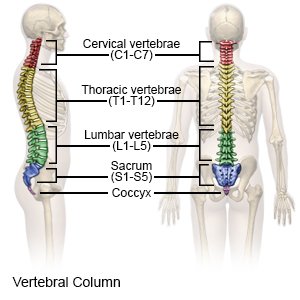
A thoracolumbar fracture is a break in a thoracic or lumbar vertebrae. The thoracic vertebrae are the 12 bones between your neck and lower back. They are connected to your ribs and help the ribs move when you breathe. The lumbar vertebrae are the 5 bones between your chest and hips. When a vertebra is damaged, the spinal cord may also be damaged.
 |
What are the signs and symptoms of a thoracolumbar fracture?
- Pain that increases during movement
- Limited movement of your back
- Abnormal curve of the spine
- Bruising and swelling on your back
- Numbness, weakness, or paralysis of your legs
How is a thoracolumbar fracture diagnosed?
- An x-ray, CT scan, or MRI will be done to check for broken bones or other problems. You may be given contrast liquid to help the bones show up better in the pictures. Tell the healthcare provider if you have ever had an allergic reaction to contrast liquid. Do not enter the MRI room with anything metal. Metal can cause serious injury. Tell the healthcare provider if you have any metal in or on your body.
- A bone scan is a test done to show areas where your bone is diseased or damaged. You will get a radioactive liquid, called a tracer, through a vein in your arm. The tracer collects in your bones and shows up in pictures.
How is a thoracolumbar fracture treated?
Treatment will depend on which bones were damaged and the kind of fracture you have.
- Bed rest may be used for a mild fracture. Bed rest helps prevent injury and protects the vertebra until it heals.
- NSAIDs , such as ibuprofen, help decrease swelling, pain, and fever. This medicine is available with or without a doctor's order. NSAIDs can cause stomach bleeding or kidney problems in certain people. If you take blood thinner medicine, always ask if NSAIDs are safe for you. Always read the medicine label and follow directions. Do not give these medicines to children younger than 6 months without direction from a healthcare provider.
- Acetaminophen decreases pain and fever. It is available without a doctor's order. Ask how much to take and how often to take it. Follow directions. Read the labels of all other medicines you are using to see if they also contain acetaminophen, or ask your doctor or pharmacist. Acetaminophen can cause liver damage if not taken correctly.
- A back brace or back cast may be used for support. Sometimes a corset (binder) may be used to support a weak spine.
- A walker may help decrease the load on a broken spine.
- Surgery may be needed for a severe fracture. Surgery is used to move the bones to their correct positions.
Call your local emergency number (911 in the US) if:
- You feel lightheaded, short of breath, or have chest pain.
- You cough up blood.
- You have trouble moving your legs.
- Your legs feel numb or you cannot move them.
When should I seek immediate care?
- Your arm or leg feels warm, tender, and painful. It may look swollen and red.
When should I call my doctor?
- You have pain or swelling on your back that is worse or does not go away.
- You have questions or concerns about your condition or care.
Care Agreement
You have the right to help plan your care. Learn about your health condition and how it may be treated. Discuss treatment options with your healthcare providers to decide what care you want to receive. You always have the right to refuse treatment. The above information is an educational aid only. It is not intended as medical advice for individual conditions or treatments. Talk to your doctor, nurse or pharmacist before following any medical regimen to see if it is safe and effective for you.© Copyright Merative 2025 Information is for End User's use only and may not be sold, redistributed or otherwise used for commercial purposes.
Learn more about Thoracolumbar Fracture
Treatment options
Symptoms and treatments
Medicine.com guides (external)
Further information
Always consult your healthcare provider to ensure the information displayed on this page applies to your personal circumstances.